 Dr. Ronald Torres Labardini ·
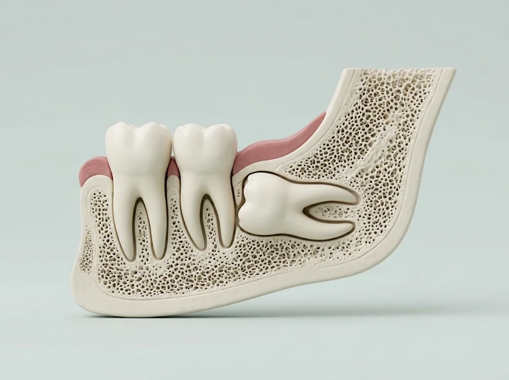
The short answer
A wisdom tooth comes out when there is a specific problem: an infection that keeps coming back, decay that can no longer be fixed, damage to the neighboring molar, or a cyst. If it's healthy and causing no trouble, the evidence isn't strong enough to say removing it is better than keeping it; in that case we monitor it with regular check-ups. I don't have one rule for everyone: we decide case by case, together. One of my 104 reviews on HuliHealth, which average 5.0, puts it this way (in Spanish): «Claridad en explicar el diagnostico y los eventuales escenarios para proceder con el tratamiento a seguir» ("clarity in explaining the diagnosis and the possible scenarios for the treatment to follow"; Maria R., April 6, 2025).
Signs it's time to remove them
These are, in essence, the situations the UK's NICE guidance considers reasons to operate on an impacted wisdom tooth:
- Pericoronitis that comes back: the gum covering the tooth gets inflamed or infected a second time or more. A first episode, unless it's severe, is usually treated without surgery.
- Decay in the wisdom tooth that can no longer be fixed, or an infection of the tooth's nerve or root tip that can't be treated.
- An abscess, cellulitis or osteomyelitis (bone infection).
- Resorption of the wisdom tooth or the neighboring molar (the tooth structure is being lost), or a fractured tooth.
- A cyst or tumor in the sac around the wisdom tooth.
- The tooth is in the way of other jaw surgery, such as a reconstruction, or lies in the area of a tumor treatment.
What if they don't hurt? The preventive-removal debate
It's a very common question. A 2020 Cochrane systematic review concluded there isn't enough evidence to decide whether impacted wisdom teeth with no symptoms and no disease should be removed or kept. The UK's NICE guidance, from 2000 and still published, goes further: it recommends against preventive removal. This is what I weigh with each patient:
- Keeping it doesn't mean forgetting it: if we decide to keep the tooth, I check it at regular intervals to catch any problem early.
- The risk of keeping it: very low-certainty evidence suggests that an impacted, symptom-free wisdom tooth may be associated, in the long term, with more gum and bone disease (periodontitis) on the neighboring molar.
- When the sac around symptom-free wisdom teeth is examined under the microscope, changes, including cystic ones, are sometimes found even when the X-ray looks normal. Even so, the evidence doesn't support removing teeth for that reason alone.
- Neither option is free of risk: when wisdom teeth lead to a hospital admission, the most reported causes are pericoronitis and infections after removal.
What the X-ray and CT scan show
This decision isn't made by eye. The panoramic X-ray shows where each wisdom tooth sits, how it's tilted, its roots, and how it relates to the inferior alveolar nerve, which gives feeling to the lip and chin. Often it's all we need.
- I look for three warning signs: the nerve canal bending away, its white outline breaking, and the root darkening where it crosses the canal. They flag a higher risk of nerve injury.
- I measure how deep the tooth is, whether it's upright, tilted or lying sideways, and whether the root touches the canal. Depth, root contact with the canal and teeth lying sideways are factors studies link to higher nerve risk.
- If those signs show up, I order a 3D CT scan (CBCT). It helps identify the risk more clearly and plan the surgery, but on its own it hasn't been shown to reduce nerve injury: its value is in helping me decide how to operate or whether another option is better.
- For perspective: depending on the study, inferior alveolar nerve injury after lower wisdom tooth removal is reported between 0.35% and 8.4%. In a review of 44,171 impacted lower wisdom tooth extractions, it was temporary in 1.20 out of 100 and did not recover in 0.28 out of 100.
Coronectomy: when the root sits against the nerve
If the root is pressed tightly against the nerve, removing the whole tooth isn't always the most appropriate option. There are two alternatives: monitoring it, or a coronectomy, where I remove only the crown and leave the roots in place, without working in the area of the nerve.
- What you gain: in observational studies, compared with full removal, it can reduce injury to the lip and tongue nerves, and dry socket as well.
- What it costs: the roots left behind may shift (13% to 85% of cases, depending on the study), and 2.2% to 3.63% needed a second surgery, depending on the review.
- In one review, the roots became exposed in 2.66 out of 100 cases and the technique failed in 2.79 out of 100.
- That's why, after a coronectomy, I follow the roots with X-rays.
At what age should they come out?
No age on its own means wisdom teeth have to come out. Age goes on the scale along with the tooth's position and your health:
- TeensIn selected cases, removing the tooth while it's still developing (germectomy) may reduce nerve risk and complications, but it comes with short-term discomfort. It isn't recommended routinely: it's decided on each person's risk.
- Over 24Being over 24 is among the factors that may increase the risk of nerve injury, along with teeth lying sideways. It's no reason to remove a healthy tooth just in case, but it's worth keeping in mind when a wisdom tooth is already causing problems.
- Any ageIf there's infection that keeps coming back, unfixable decay, damage to the neighboring molar or a cyst, the indication is the same. And if the tooth is healthy, monitoring is an option for adults too.
What to bring to your consultation
- If you already have a panoramic X-ray or a CT scan, bring it or send it ahead: it avoids repeating imaging, and we can review it first by video.
- A list of your medications and allergies. If you take blood thinners, don't stop them on your own: I coordinate with your doctor.
- Your questions. A good one to start with: what happens if I keep it?
- A consultation is 60,000 colones at my office in Tower 1 of Hospital CIMA, and 20,000 by video. I give you the cost of surgery after reviewing your imaging.
When to message me or go to the ER
- Message me or call the office if the gum behind your last molar swells, hurts or bleeds, or if you notice a bad taste or smell there: it may be pericoronitis.
- If you've already had surgery: severe pain that starts between day 1 and day 5, sometimes with bad breath, may be dry socket. Message me: it's treated at the office.
- A fever or swelling that keeps growing: call the office right away; if you can't reach me, go to the ER.
- Go to the ER right away if swelling spreads toward your eye, neck or under your tongue, or if you have trouble swallowing, breathing or opening your mouth.
Sources
- 1.Ghaeminia H et al. — Surgical removal versus retention for the management of asymptomatic disease-free impacted wisdom teeth. Cochrane Database Syst Rev, 2020.
- 2.NICE — Guidance on the extraction of wisdom teeth (technology appraisal guidance TA1). National Institute for Health and Care Excellence, UK, 2000.
- 3.de Andrade Tutu JS et al. — Cystic and Neoplastic Lesions in Pericoronal Follicles of Asymptomatic Third Molars: A Systematic Review and Meta-Analysis. J Oral Maxillofac Surg, 2026.
- 4.Lau T et al. — Potentially Preventable Hospitalisation Related to Severe Third Molar Disease: A Systematic Review. Dent J (Basel), 2026.
- 5.Su N et al. — Predictive Value of Panoramic Radiography for Injury of Inferior Alveolar Nerve After Mandibular Third Molar Surgery. J Oral Maxillofac Surg, 2017.
- 6.Kang F et al. — Determining the risk relationship associated with inferior alveolar nerve injury following removal of mandibular third molar teeth: A systematic review. J Stomatol Oral Maxillofac Surg, 2020.
- 7.Sarikov R, Juodzbalys G — Inferior alveolar nerve injury after mandibular third molar extraction: a literature review. J Oral Maxillofac Res, 2014.
- 8.Clé-Ovejero A et al. — Does 3-dimensional imaging of the third molar reduce the risk of experiencing inferior alveolar nerve injury owing to extraction?: A meta-analysis. J Am Dent Assoc, 2017.
- 9.Ghaeminia H et al. — Clinical relevance of cone beam computed tomography in mandibular third molar removal: A multicentre, randomised, controlled trial. J Craniomaxillofac Surg, 2015.
- 10.Kang FW et al. — Coronectomy in Lower Third Molar Surgery: A Systematic Review and Meta-Analysis. J Oral Maxillofac Surg, 2025.
- 11.Pitros P et al. — A systematic review of the complications of high-risk third molar removal and coronectomy: development of a decision tree model and preliminary health economic analysis to assist in treatment planning. Br J Oral Maxillofac Surg, 2020.
- 12.Scilla F et al. — Lower Third Molar Germectomy: Timings, Indications, and Clinical and Patient-Reported Outcomes-A Systematic Review and Meta-Analysis. Dent J (Basel), 2026.
- 13.Kuśnierek W et al. — Smoking as a Risk Factor for Dry Socket: A Systematic Review. Dent J (Basel), 2022.