08 · Oral & maxillofacial surgery
Jaw cysts and tumors: diagnosis and surgery
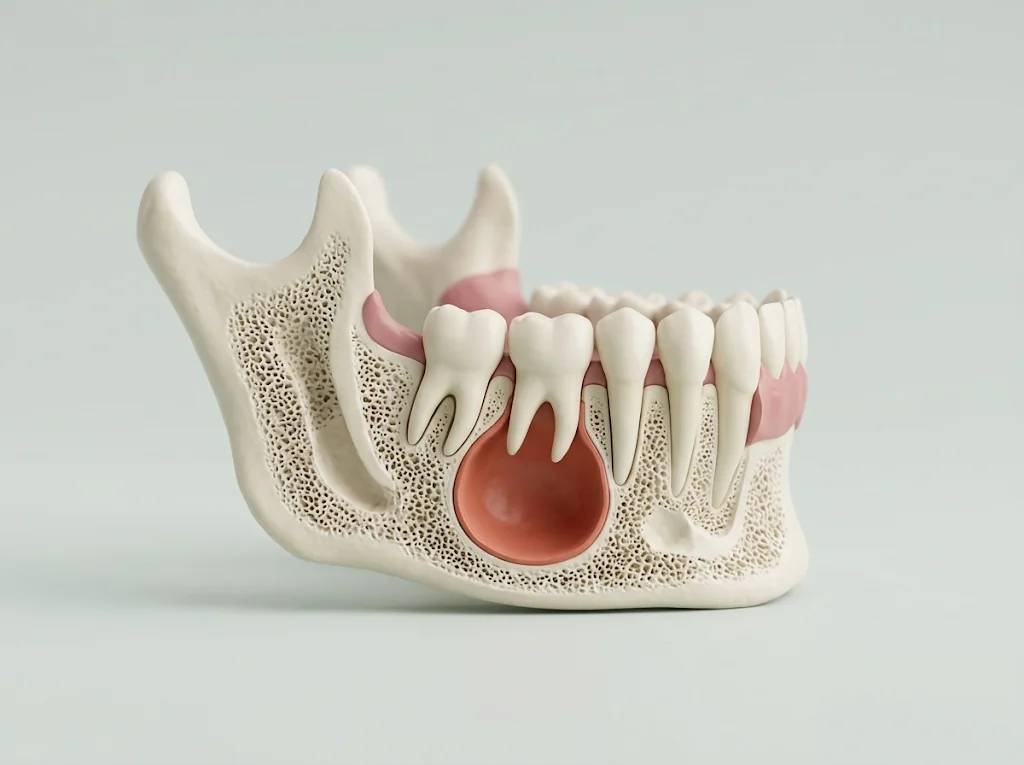
Many jaw cysts don't hurt: they show up on a routine X-ray or as a bump on the gum. They still need treatment, because they grow and thin the bone. Before I operate I measure the lesion on a CT scan and confirm what it is with a biopsy; with that diagnosis I walk you through the options and we decide together.
Book a visit soon if you have an unexplained ulcer lasting more than 3 weeks, a red patch, or a lump in your neck that won't go away. If your face swells quickly, you have a fever, or it's hard to swallow or breathe, go to the emergency room.
- Imaging
- Panoramic X-ray and CT scan (CBCT)
- Diagnosis
- Biopsy and pathology
- Anesthesia
- Local; for larger lesions, per the plan
- Follow-up
- Clinical and with X-rays
When it's indicated
- A dark area or a cyst in the bone on a routine X-ray.
- A swelling of the gum, palate or jaw, or a tooth that shifts for no reason.
- A cyst around a wisdom tooth or canine that never came in (dentigerous cyst).
- An unexplained ulcer lasting more than 3 weeks, or a sore that hasn't healed 2 weeks after removing whatever was rubbing it.
- A red patch, a red-and-white patch, or a white one that doesn't rub off.
- A papilloma or another lump on the lip or inside the mouth.
Before surgery
- Bring any X-rays or CT scans you have, with their reports: sometimes they spare you a repeat scan.
- List your medications. If you take blood thinners, don't stop them on your own: continuing is usually preferred, because stopping is associated with a higher risk of clots. I coordinate it with your doctor.
- If there is sedation or general anesthesia, I give you written instructions on fasting and on bringing a companion.
- If the result scares you, tell me. Anxiety can make people put off the diagnosis, which is why the American Dental Association guideline stresses talking it through.
How I plan it
How I do it
- 01AssessmentExamination and imaging. If a mouth lesion may come from rubbing (a sharp tooth, a denture), we remove the cause first and recheck it at 2 weeks.
- 02BiopsyUnder local anesthesia, with a few stitches. The sample goes to the pathologist; results take from days to a few weeks, and I go over them with you calmly.
- 03SurgeryDepending on the diagnosis: enucleation (removing the whole cyst), sometimes with cleaning of the surrounding bone or Carnoy's solution; for large cysts, decompression first — a small opening so the cyst shrinks — and removal later; for ameloblastoma, resection with margins or, in selected cases, conservative treatment.
- 04Pathology of what I removeEverything I remove goes to the pathologist, to confirm the diagnosis and that it came out completely. I remove a papilloma with a margin of healthy tissue: in one case series, it came back less often with at least 3 mm.
- 05Follow-upX-rays to confirm the bone fills in and the lesion doesn't return. For keratocysts and ameloblastomas, for years.
Recovery
- Biopsy: mild discomfort for a few days, soft food and the prescribed medication.
- Cyst: swelling is greatest in the first days and then goes down. Cold packs, a soft diet and a visit to check the area and the stitches.
- If the bone was left thin, a soft diet for longer. It fills in gradually; we follow it with X-rays.
- With decompression, the process usually lasts one to two years and generally ends with a second, less invasive surgery.
- A white or red patch that turned out not to be cancer still needs periodic, long-term follow-up.
Risks and how I prevent them
- The cyst coming back
- This is the main risk with keratocysts, which tend to recur: published figures range from 3% to 60% depending on the study. With enucleation (alone or with curettage, depending on the review), 22.5% to 23.1%; adding Carnoy's solution, 5.3% to 11.5%. I manage it by removing the lesion completely, with whatever adjunct the case calls for, and with follow-up.
- The ameloblastoma coming back
- In the solid or multicystic type it came back in 8% after bone resection and in 41% after conservative treatment; in the unicystic type, in 3% and 21%. These data come only from retrospective studies. If we choose the conservative route, check-ups are more frequent.
- Numbness of the lip or chin
- It can happen when the lesion surrounds the inferior alveolar nerve. That's why I measure that distance on the CT scan and plan the approach to protect it.
- Neighboring teeth and fracture
- A large cyst can involve nearby roots, which sometimes need a root canal or have to come out. If it thins the bone a lot, there is a risk of fracture: decompressing first may be the better choice.
- Bleeding and infection
- As with any surgery. With direct oral anticoagulants, bleeding after oral procedures was similar whether the drug was continued (11.8%) or stopped (13.5%), and all bleeding was controlled with local measures.
Other options
- Decompressing first
- For large cysts, a small opening shrinks them over one to two years and makes the second surgery less invasive. It hasn't been firmly shown to lower recurrence compared with enucleation alone.
- Resecting the bone
- For keratocysts it had the lowest recurrence in two reviews, 2.2% to 8.4%, but with a bigger impact. The authors of one propose reserving it for lesions that recur several times and, possibly, syndromic cases.
- Waiting and watching
- Reasonable for a typical canker sore, which heals on its own in one to two weeks (a topical steroid eases the pain and shortens healing), or for a sore whose cause gets corrected. Not for a bone lesion without a diagnosis.
Frequently asked questions
How is a jaw cyst found?
Often by chance, on an X-ray taken for another reason; other times because of a bump on the gum or a tooth that shifts. Many don't hurt.
Is a cyst in the jaw cancer?
Generally, no: keratocysts and ameloblastomas are benign, although they can grow and come back. The X-ray points the way; what confirms what it is — and rules out cancer — is the biopsy. In 2009 I took the oral and maxillofacial oncology course at Mexico's National Cancer Institute.
Does a cyst always need surgery?
A diagnosed cyst is treated, because it usually keeps growing. What changes is how, depending on the type and size. What I don't recommend is watching a bone lesion without a biopsy.
What is a dentigerous cyst?
A cyst that forms around the crown of a tooth that never came in, often a wisdom tooth or a canine. Depending on the case, it comes out with the tooth or is decompressed first.
What is an ameloblastoma?
A benign tumor that starts in the tissues that form teeth. It isn't cancer, but it comes back more often after conservative treatment than after bone resection; treatment is decided by the biopsy, the type and the size.
Can the cyst come back after surgery?
It depends on the type. In an analysis of 6427 keratocysts, overall recurrence was 21.1%: 14.7% in single-cavity lesions, 24.4% in multi-cavity ones and 35.4% in Gorlin syndrome. That's why X-ray check-ups go on for years.
Does a biopsy hurt, and when are the results ready?
Under local anesthesia, you usually feel pressure; if anything hurts, I add more anesthetic. Afterwards, mild discomfort for a few days. Results take from days to a few weeks; if easier, I can go over them by video consultation.
Sources
- 1.NICE — Suspected cancer: recognition and referral (NG12). Recommendations organised by site of cancer. National Institute for Health and Care Excellence, 2015.
- 2.Abati S et al. — Oral Cancer and Precancer: A Narrative Review on the Relevance of Early Diagnosis. Int J Environ Res Public Health, 2020.
- 3.Engelen ET et al. — Antifibrinolytic therapy for preventing oral bleeding in people on anticoagulants undergoing minor oral surgery or dental extractions. Cochrane Database Syst Rev, 2018.
- 4.Lingen MW et al. — Evidence-based clinical practice guideline for the evaluation of potentially malignant disorders in the oral cavity: A report of the American Dental Association. J Am Dent Assoc, 2017.
- 5.Sharif FN et al. — Interventions for the treatment of keratocystic odontogenic tumours. Cochrane Database Syst Rev, 2015.
- 6.Nammour S et al. — Success Rate of Benign Oral Squamous Papilloma Treatments After Different Surgical Protocols (Conventional, Nd:YAG, CO2 and Diode 980 nm Lasers): A 34-Year Retrospective Study. Photobiomodul Photomed Laser Surg, 2021.
- 7.Berretta LM et al. — Effectiveness of marsupialisation and decompression on the reduction of cystic jaw lesions: a systematic review. Br J Oral Maxillofac Surg, 2021.
- 8.Iocca O et al. — Potentially malignant disorders of the oral cavity and oral dysplasia: A systematic review and meta-analysis of malignant transformation rate by subtype. Head Neck, 2020.
- 9.Al-Moraissi EA et al. — What surgical treatment has the lowest recurrence rate following the management of keratocystic odontogenic tumor?: A large systematic review and meta-analysis. J Craniomaxillofac Surg, 2017.
- 10.Chrcanovic BR, Gomez RS — Recurrence probability for keratocystic odontogenic tumors: An analysis of 6427 cases. J Craniomaxillofac Surg, 2017.
- 11.Hendra FN et al. — Radical vs conservative treatment of intraosseous ameloblastoma: Systematic review and meta-analysis. Oral Dis, 2019.
- 12.Gasparro R et al. — The Effect of Conservative vs. Radical Treatment of Ameloblastoma on Recurrence Rate and Quality of Life: An Umbrella Review. J Clin Med, 2024.
- 13.Manfredi M et al. — World workshop on oral medicine VII: Direct anticoagulant agents management for invasive oral procedures: A systematic review and meta-analysis. Oral Dis, 2019.
- 14.Slusarenko da Silva Y et al. — Recurrence of nonsyndromic odontogenic keratocyst after marsupialization and delayed enucleation vs. enucleation alone: a systematic review and meta-analysis. Oral Maxillofac Surg, 2019.
- 15.Le Joncour A et al. — [Idiopathic and secondary recurrent aphthous stomatitis: A narrative review]. Rev Med Interne, 2026.
- 16.Al-Aizari NA et al. — Evidence-based recommendations for the treatment of recurrent aphthous stomatitis: insights from an umbrella review. J Dermatolog Treat, 2026.
Reviews from my patients (not necessarily about this procedure)
«Desde que entramos al área de parqueo se siente la seguridad y confianza, la recepcionista muy amable y simpática, poco antes de la hora de la cita nos avisan que había un pequeño atraso por una complicación con el paciente que tenía el Dr. Torres en ese momento la espera no fue larga y de todos modos el área de espera es muy amplia y agradable, El Dr. Torres muy amable y profesional en todo momento, no escatimó tiempo para dar una amplia explicación del procedimiento, en general mi esposa y yo nos sentimos muy cómodos y sobretodo con mucha confianza de estar en las manos adecuadas.Definitivamente 100% recomendados !»«El Doctor Ronald Torres es un excelente profesional. Lo recomiendo 100%. Su trato siempre ha sido amable, cortes, profesional. Se interesa en comprender la necesidad del paciente, y en todo momento trata que este comprenda bien los alcances del procedimiento a realizar. Ya me ha extraido dos cordales, y puedo decir que me fue muy bien. Puede tener confianza de buscar al Dr. Ronald, que le aseguro quedara satisfecho con su atencion.»«Excelente profesional, impecable en la técnica de su especialidad y con un trato humano digno de resaltar. Desde el día uno nos sentimos cómodos con él y esto se mantuvo y se ha mantenido hasta el día de hoy, en donde estamos ya con citas de mero seguimiento. La intervención quirúrgica realizada fue un éxito y el costo muy razonable. La verdad que gracias a Dios tomamos una excelente decisión con él. Súper recomendado.»«Mi hijo Luis Guillermo se realizó una cirugía maxilofacial con el doctor Torres y su equipo, el resultado fue y es a hoy excelente, recibió un servicio excelente de seguimiento después de su cirugía.Le cambió mucho la vida a mi hijo de forma positiva , una gran inyección de seguridad y autoestima.Como padres nos sentimos muy agradecidos y es de esas inversiones que valen la pena .»Other procedures
- 01Wisdom teeth surgery
- 02Dental implants
- 03All-on-4 immediate full arch
- 04Orthognathic surgery
- 05Sinus lift
- 06TMJ treatment and surgery
- 07Facial trauma
- 09Buccal fat removal
- 10Extractions and abscesses
- 11Bone grafting and socket preservation
- 12Oral lesions and biopsy
- 13Zygomatic implants
- 14Video consultation and second opinion